After treatment
A feeling of pressure or tenderness when biting may occur for a few days. Do not eat again until the local anesthesia has completely worn off.
Endodontics · Berlin-Mitte
Magnification and light aligned with the line of sight help identify fine canal structures and treat them precisely. This improves the working conditions but does not guarantee success.
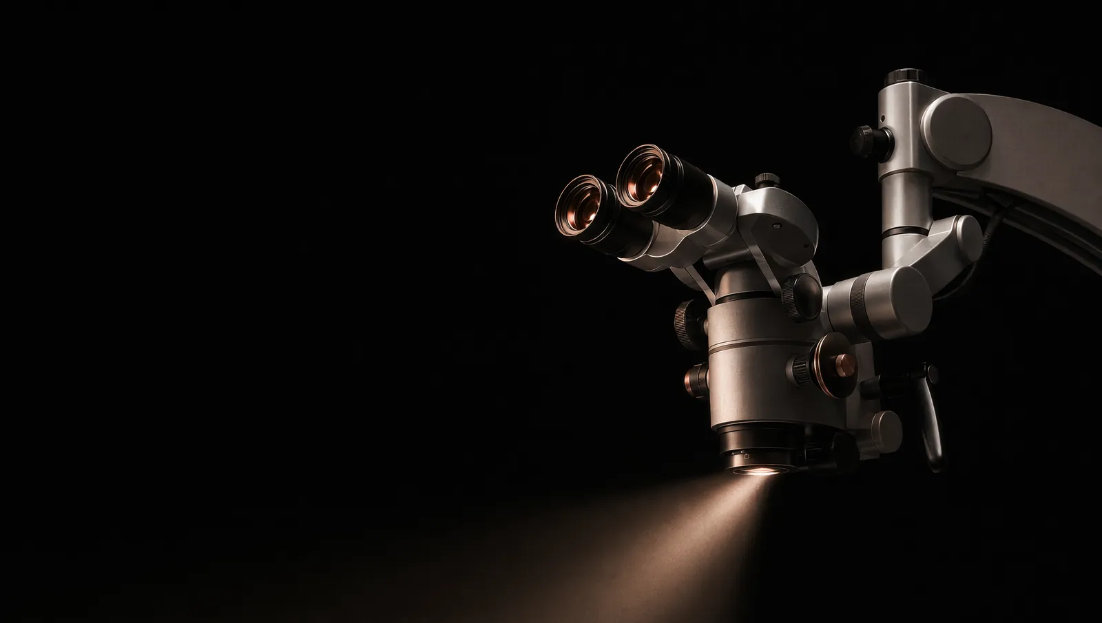
Microscope
Inside the tooth is the pulp, a tissue made up of nerves and blood vessels. If it is irreversibly inflamed or has died, the tissue and bacteria are removed from the branching canal system. The canals are then disinfected and filled, and the tooth is tightly sealed.
A dental microscope combines magnification with coaxial light. The light is directed deep into the tooth along the line of sight. This helps identify additional canals, navigate narrow or calcified access points, and assess teeth that have already been treated.
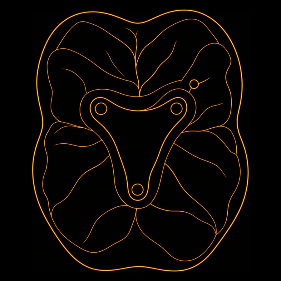
The first upper molar often has a second canal in the mesiobuccal root, known as MB2. In an international study of 5,250 first maxillary molars, it was found in 73.8 percent of the teeth examined.
This figure shows why looking for additional anatomy is relevant. It does not predict how many canals your tooth has. We determine that from your individual findings.
Source: International multicenter CBCT study.
Better visibility does not solve every problem. Deep cracks, inaccessible anatomy, or insufficient remaining tooth structure can limit whether the tooth can be preserved despite the microscope.
Guidance
Whether an initial root canal treatment, another attempt to preserve the tooth, or a different restoration is appropriate depends on the tooth's prognosis. These three questions show what we examine and discuss first.
How has the tooth been treated so far?
What have you been told about the prognosis?
What would you like to clarify during the consultation?
If the tooth appears restorable and has not yet had root canal treatment, we discuss initial treatment under the microscope. Preserving the tooth requires a treatable canal system and sound remaining tooth structure. An examination and X-ray clarify the starting point.
Planning also includes a tight seal and the tooth's subsequent stable restoration.
For a tooth that has already been treated, retreatment under the microscope can provide access to existing filling material and structures that were not previously reached. Retreatment and apicoectomy address different clinical questions. Which option is appropriate depends on the anatomy, the cause, and whether the tooth can be restored.
Before making a decision, we review existing images, the root filling, the remaining tooth structure, and the previous restoration.
If there may be a crack, little remaining tooth structure, or an unclear periodontal prognosis, no treatment has yet been decided. The findings determine whether an attempt to preserve the tooth is clinically justifiable. Only then do we compare root canal treatment, extraction, and possible replacement.
If replacement is an option, the bone, neighboring teeth, prosthetic replacement, and follow-up care must be considered together.
These questions do not replace an examination, diagnosis, or consultation. They only help indicate which treatment pathway should be assessed. Which option may be suitable for your tooth depends on the findings and prognosis.
Process
The steps follow an established clinical sequence. How much can be done in one session depends on the tooth, the infection, and the existing restoration.
An examination, pulp sensibility tests, and digital X-rays allow us to determine whether the pulp is irreversibly inflamed or has died and whether the tooth can be preserved. Before treatment begins, we explain the available alternatives.
Treatment is performed under local anesthesia. A rubber dam, a flexible sheet placed around the tooth, keeps saliva and bacteria away. It also protects you from irrigating solutions and small instruments and is standard at F80.
The access opening to the canal system is prepared under magnification and coaxial light. Canal openings, existing filling materials, calcifications, and anatomical features are assessed under direct vision.
The working length of each canal is measured electronically. This determines how far the canal should be cleaned and filled. If medically necessary, an X-ray check supplements the measurement.
Flexible nickel-titanium instruments mechanically shape the canals. The aim is to create a controlled space for disinfection and filling without removing more tooth structure than necessary.
Disinfecting irrigants reach areas that an instrument does not touch. The protocol is based on the anatomy and the extent of infection. For selected findings, laser application may supplement disinfection; it does not replace mechanical cleaning.
Obturation is the filling of the cleaned canals. They are sealed with gutta-percha and a sealing material. This is followed by a tight coronal seal, meaning closure of the tooth above the roots. The result is checked.
Depending on the defect, the tooth receives a durable filling, an inlay, a partial crown, or a crown. This definitive restoration is part of the prognosis: a severely weakened root canal-treated tooth is more prone to fracture. At follow-up appointments, we check the healing process.
Sessions
Giving a blanket number would not be responsible. A finding that is readily accessible and can be controlled may be treated in one session. In cases of pronounced infection, fluid discharge, complex anatomy, or retreatment, an intracanal medication and another appointment may be appropriate.
A feeling of pressure or tenderness when biting may occur for a few days. Do not eat again until the local anesthesia has completely worn off.
Increasing or severe symptoms, swelling, fever, or a lost temporary seal should be assessed promptly.
They are not routinely part of root canal treatment. A clear indication may exist in cases such as fever, a general feeling of illness, or a spreading infection.
Antibiotics replace neither the opening and cleaning of the canal system nor necessary drainage. Medication and pain relief are discussed individually based on your medical history.
Prognosis
A systematic review found success rates for nonsurgical primary root canal treatments of 82.0 percent under strict criteria and 92.6 percent under less strict criteria.
Systematic review of primary root canal treatments – PubMed 35334111. These are group values from studies, not an F80 success rate and not a promise for an individual tooth. Initial findings, anatomy, disinfection, seal integrity, remaining tooth structure and the definitive restoration affect the individual prognosis.
A natural tooth that can be preserved is biologically valuable. Root canal treatment avoids extraction but does not heal every tooth. A fracture that cannot be restored, insufficient sound remaining tooth structure, an infection that cannot be controlled, or a very poor periodontal prognosis may weigh against preserving the tooth.
An implant replaces a lost tooth and requires a surgical procedure as well as separate planning for bone and soft tissue. It is not categorically better or worse. We compare the prognosis, treatment effort, follow-up care, and private services for both pathways. More information about replacing a tooth that cannot be preserved is available under Dental implants.
A treatable canal system, sufficient sound remaining tooth structure, and an acceptable periodontal prognosis support preserving the tooth. The existing tooth remains in the jaw. It then needs a tightly sealed core build-up, a partial crown or crown depending on the defect, and further checkups.
A fracture that cannot be restored, an inaccessible infection, or insufficient load-bearing tooth structure may weigh against preserving the tooth.
An implant replaces the root of the extracted tooth. This requires an assessment of the available bone, soft tissue, and general health. The surgical procedure is followed by a healing period, restoration with a crown, and ongoing implant follow-up care.
Switching to an implant does not automatically improve quality. It is a separate treatment with its own requirements and risks.
Costs
The gesetzliche Krankenkasse (German statutory health insurance) covers root canal treatment under certain conditions. The tooth must be capable of preservation and clinically worth preserving. It must be possible to prepare and fill the canals to or near the root tip.
Additional criteria apply to molars. For example, treatment should preserve an uninterrupted dental arch, avoid a unilateral free-end situation, or preserve an existing functional dental prosthesis. We determine from the findings whether these conditions are met.
Additional microscope-assisted services and other procedures often require private payment. Before treatment begins, you receive a written plan that separates services covered by the gesetzliche Krankenkasse (German statutory health insurance) from private components.
Treatment

Dentist · Endodontics
Ly Cao is a dentist specialising in endodontics. She studied dentistry at the University of Greifswald and completed the DGET endodontology curriculum in 2020.
She provides treatment in German, English, and Vietnamese.
Questions
It is indicated when the tissue inside the tooth is irreversibly inflamed or has died and the tooth can be preserved. The canal system is cleaned, disinfected, and tightly filled.
Treatment is performed under local anesthesia. You may feel pressure, movement, or the effects of keeping your mouth open for an extended period. If you feel pain, tell us immediately; treatment will be paused and the anesthesia checked.
No. It improves illumination and visibility of additional canals, calcifications, and complex findings. The prognosis also depends on the infection, anatomy, disinfection, tight seal, remaining tooth structure, and definitive restoration.
One or more sessions may be clinically appropriate. The determining factors are the tooth, the number and shape of the canals, the extent of infection, isolation, and any previous treatment. A fixed number cannot be promised before diagnosis.
The rubber sheet keeps saliva and bacteria away from the opened tooth. At the same time, it protects you from irrigating solutions and small instruments. At F80, it is part of root canal treatment.
A feeling of pressure and tenderness when biting may occur for a few days. Please contact us promptly if you have severe or increasing symptoms, swelling, fever, or if a temporary restoration is lost.
Not routinely. Antibiotics may be considered when there is a clear indication, such as fever, a general feeling of illness, or a spreading infection. The cause inside the tooth must still be treated.
Not every tooth needs the same restoration. If a large amount of tooth structure is missing, the tooth is more prone to fracture. A partial crown or crown may stabilize it. For smaller defects, a durable filling or an inlay may be sufficient.
The gesetzliche Krankenkasse (German statutory health insurance) covers root canal treatment if its guidelines for the affected tooth are met. Additional microscope-assisted services often require private payment. You receive a written plan before treatment begins.
Not in general. A tooth that can be preserved may function for a long time after root canal treatment and a stable restoration. If it cannot be restored or has a very poor prognosis, an implant may be more appropriate. We compare both pathways based on the findings.
At Friedrichstraße 80, we determine whether the tooth can be preserved, how complex the canal system is, and which services are planned. You decide on this basis.
Or call: 030 436665501